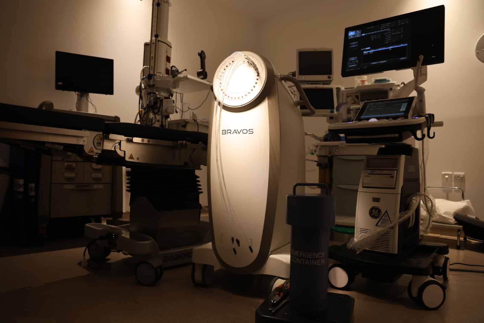
What do you know about radioactive seed therapy?
Article: Iqbal Al-Amriy – Medical Physicist
Brachy radiotherapy is a special form of radiotherapy known as brachy, which means short or near, referring to the close distance between the radiation source and the tumor. In contrast to usual external radiotherapy, the potent radioactive source of radiation is placed directly on or inserted into the tumor. This allows for targeted and precise irradiation of the tumor area, providing the best possible protection for surrounding healthy body tissue. Brachytherapy may be used as monotherapy or in combination with external radiation therapy, depending on the indication.
In proximity radiotherapy, as in almost all forms of radiotherapy, gamma rays produced by the natural decay of atomic nuclei are used. The potent radioactive radiation from this causes damage to the body’s cells, and the very rapid growth of many cancer cells makes them more vulnerable to radiation than the surrounding tissues.
There are two types of radiation therapy closely:
- Permanent implantation, also known as low-dose brachytherapy (LDR): The radiation source is permanently implanted into the tumor. The radiation source (radioactive seeds, which are iodine-125 particles) is as small as a sesame seed. A very small amount of low-level radiation is released slowly until it is exhausted. . This type of radiotherapy is often used in close proximity to treat low-risk localized prostate cancer.
- Temporary implantation, also known as high-dose brachytherapy (HDR): A radiation source (miniature Iridium 192 sources) is implanted directly into or near the tumor temporarily where a high-level dose of radiation is released. This type of proximity radiotherapy is most often used to treat cervical cancer but can also be used for other cancers such as lung cancer, breast cancer, prostate cancer, head and neck cancer and anal cancer.
Like any type of radiotherapy, here too one begins with planning the procedure, and this requires certain information. An accurate knowledge of the extent, type and location of the tumor is essential for this. Therefore, different diagnostic procedures must initially be used, and this includes, among others: x-rays, computed tomography, magnetic resonance imaging and ultrasound. At the same time, the radiation therapist draws up a treatment plan, which includes, among other things: the size of the radiation dose, the duration of its effect, and the number of sessions that will be required.
With the help of data obtained through radiography, modern data processing can produce 3D images of the tumor with its exact location in the body. In special programs, it is then possible to plan the optimal distribution of radiation sources and calculate radiation doses that affect the tumor and surrounding tissue.
If this is done successfully, the next step can be taken, which is the insertion of the applicators: the applicator is the delivery system for the radioactive active material that is subsequently inserted, and using the previously calculated image data, the exact position of the applicators in the body, where they are then fixed, can be reached. And all these steps are done on the same day for one session, and then the patient can leave and go about his life normally.


